Wednesday, April 05, 2006

Opus Princeps of Trans-Iridial Studies : Data

by Dr Dan Waniek, MD
Copyright © 2006 danwaniek.org

We report here a simple observation to support our functional model : a spatial correspondence between :

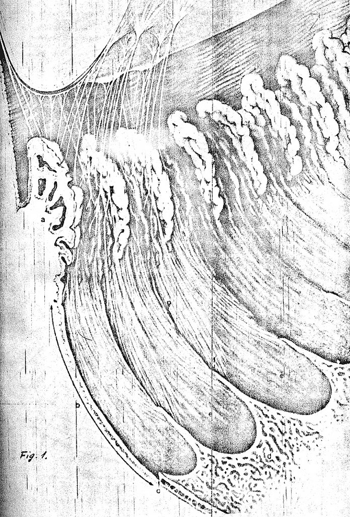
(a) radial transparencies in iridial structure,
(b) circular sectors in iridological charts,
(c) inter-processural spaces ( the ciliary valleys ) and
(d) processi dentati,

does indeed exist ( Fig. 3 ).


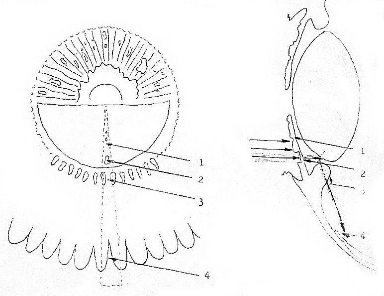
Fig. 3 : A novel observation concerning the anterior pole's peripherally aligned ( APPA ) vertebrate ocular structures : The spatial correspondence between the radial ( meridional ) structural elements of the iris, the lens and the peripheral retina, as they are viewed from the interior of the eye ( left ) and in medio-sagittal section ( right ) is put into evidence and more diagrammatically shown. There are radial transparencies ( 1 ) and stromal crypts ( 2 ) in the stroma of the iris, and these structures are in a good alignment with the ciliary valleys ( the free space between two adjacent processi ciliares ( 3 ) in pars plicata ). This alignment is shown further in pars plana, where the dentate processi of the retina ( 4 ) protrude along the symmetry axes provided by the striae ( which are not shown ). Light is permitted to reach the processi dentati along this path, considering that total reflection takes place on the anterior surface of the lens and its inter-strata. Only the inferior half of the lens is figured in the left panel. The dots at left stress the repetitive functional unit described here.

Copyright © 1986-2006 danwaniek.org ( Digimarc creator ID 10-727074 ) and the Iris-Ward.com, Inc. Based on an illustration originally published by Churchill Livingstone, with permission. All Rights Reserved.

Opus Princeps of Trans-Iridial Studies : Hypotheses

by Dr Dan Waniek, MD
Copyright © 2006 danwaniek.org

We have hypothesized that the iris may be able to transmit light through its stroma in spite of the irregularly dispersed, heavily absorptive pigment. This transmitted light, without interfering with the centro-retinal image because of total reflection on the anterior surface of the lens is, however, significant, and acts as a coded non-visual stimulus for the periphery of the retina.

The peripheral retina consists of a highly regular array of about seventy protrusions of nervous tissue, the processi dentati, in the pars plana of the ciliary body, collectively named the ora serrata.

Yet Another Functional Orphan : Peripheral Retina

This structure has not been understood so far in terms of vision : Indeed, campimetry fails to demonstrate correspondent protrusions in the margins of the visual field, even if there is positive histological evidence of normal receptive retinal structure in the ora serrata, to account for visual perception.

In our model the ora serrata is assumed to have a previously undescribed non-visual function. A critical test of these hypotheses would be to determine whether electro-retinography (ERG) can detect specific activity in the ora serrata when the surface of the iris, and not the pupil is light-stimulated.

This study was undertaken because of controversy existing between the conclusions of different approaches concerning the evaluation of iridology, the controversial diagnostic method based on clinical and empirical studies of the iridial morphology.

Opus Princeps of Trans-Iridial Studies : Introduction

by Dr Dan Waniek, MD
Copyright © 2006 danwaniek.org

The obvious function of the iris is to provide an optical diaphragm for the system that produces images on the central part of the retina.

More Detail From the Interior of the Eye

Interestingly, the morphology of the iridial stroma and epithelia seem to support more than the necessities of a highly aperture-adjustable structure. Its crypts and chromatic accidents offer a variable and complicated pattern of densities and transparencies that have stimulated several studies, both scientific and empirical.

Novel Trans-Iridial Functional Approach